A newly identified transitional state helps endothelial cells adapt to mechanical stress while preserving vascular integrity.
Jonas Franz from the research group led by Professor Dr. em. Hans Schnittler (Institute of Anatomy and Vascular Biology, University of Münster) as well as participating scientists were honored with the Quarterly Publication Award (QPA) I/2026 from the Anatomische Gesellschaft for their study, VE-Cadherin-Actin Regulation Promotes Mechanotransduction and Monolayer Maturation Involving a Tension-Sensitive Intermediate State which was recently published in the renowned journal Advanced Science.
First author as well as the lead and corresponding author of the award-winning study: Jonas Franz (left) and Professor Dr. em. Hans Schnittler (right) from the Institute of Anatomy and Vascular Biology at the University of Münster, Germany (Photos: courtesy of the authors).
Endothelial cell layers maintain tissue homeostasis by balancing structural stability with the ability to adapt to physiological and mechanical challenges. The study identifies, for the first time, a shear stress-induced, tension-sensitive (actomyosin-mediated) intermediate state as a key transitional phase on the path toward an arterial endothelial phenotype. This state stabilizes both the cell and cell-cell junctions, transiently enhances barrier function and exerts a cytoprotective effect that enables a precise and controlled reorganization of endothelial cells. These findings establish endothelial remodeling as an actively regulated, mechanosensitive process of cellular plasticity.
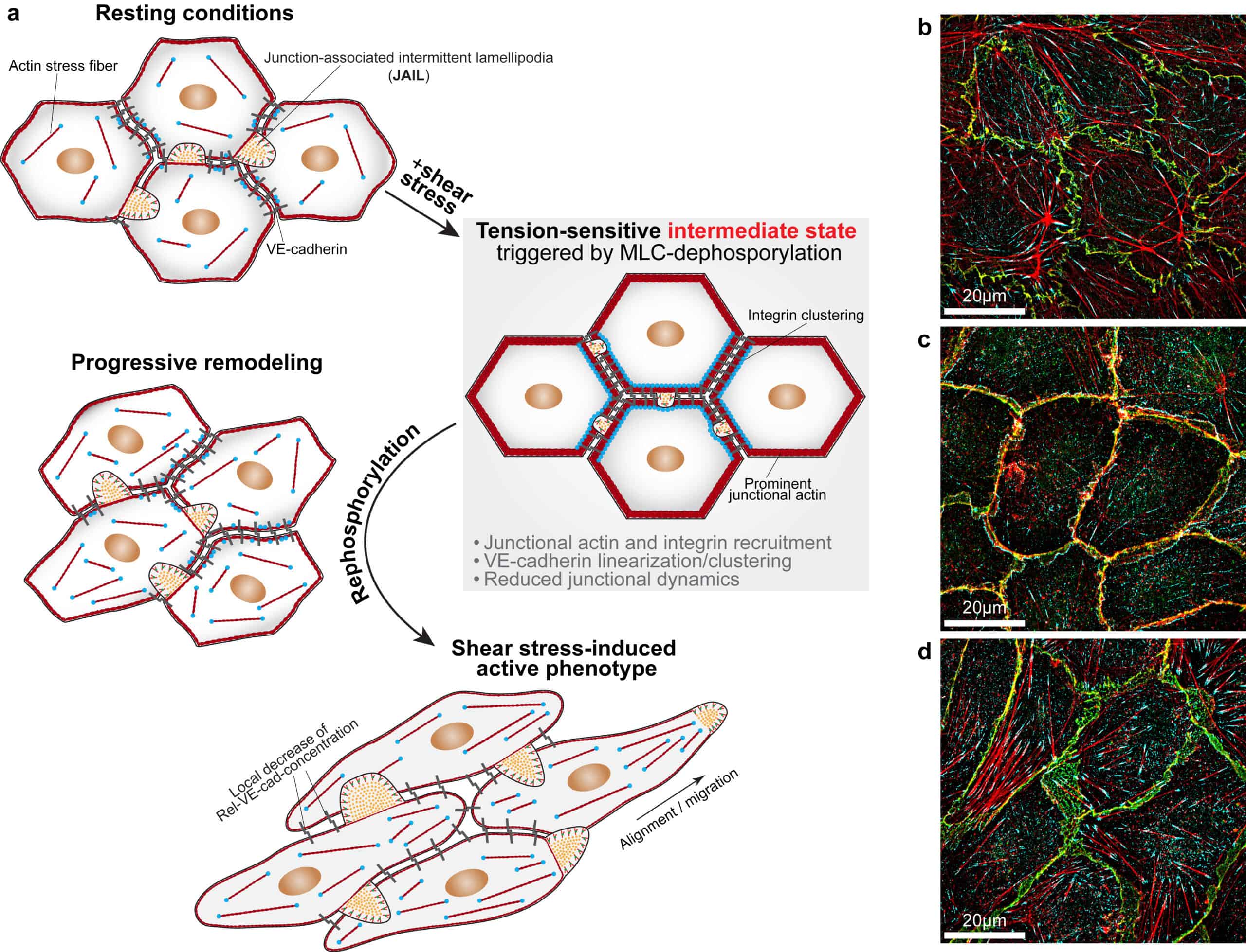
A tension-sensitive intermediate state mediates shear stress-induced endothelial remodeling and activation. (a) Scheme illustrating shear stress-induced endothelial remodeling from a resting to an active state: Resting endothelial cells (ECs) exhibited cell-cell junctions characterized by moderate junctional actin organization, high local VE-cadherin concentration and low junctional dynamics, as reflected by small junction-associated intermittent lamellipodia (JAIL). Upon 20 minutes of shear stress exposure, cells rapidly entered a transient, tension-sensitive intermediate state marked by myosin light chain (MLC) dephosphorylation, recruitment of prominent junctional actin and integrins, enhanced VE-cadherin clustering and linearization, increased barrier function and further suppression of JAIL formation. Consequently, cells adopted a homogeneous cobblestone-like morphology. Subsequent MLC-rephosphorylation initiated endothelial remodeling by dissolving junctional actin, promoting stress fiber reformation and VE-cadherin dilution, increasing JAIL-mediated junctional dynamics and weakening barrier function. These changes facilitated cell shape adaptation and migration, ultimately leading to the characteristic elongated phenotype aligned with the direction of flow. (b-d) Merged structured illumination microscopy images of ECs under (b) resting conditions, (c) during the tension-sensitive intermediate state and (d) after 4 hours of shear stress exposure. Cells were labelled for VE-cadherin (green), integrin b1 (cyan) and actin filaments (red). Panels (a-d) were assembled and modified from data published by Franz et al., 2026. Adapted under the terms of the Creative Commons Attribution 4.0 International License (CC BY 4.0).