Neue Erkenntnisse zum Remodeling von Endothelzellen
Ein neu identifizierter Übergangszustand hilft Endothelzellen, sich an mechanische Belastungen anzupassen.
Jonas Franz aus der Arbeitsgruppe von Professor Dr. em. Hans Schnittler (Institut für Anatomie und Vaskuläre Biologie der Universität Münster) sowie weitere beteiligte Wissenschaftlerinnen und Wissenschaftler wurden für Ihre kürzlich in der renommierten Fachzeitschrift Advanced Science erschienene Studie, VE-Cadherin-Actin Regulation Promotes Mechanotransduction and Monolayer Maturation Involving a Tension-Sensitive Intermediate State mit dem Quarterly Publication Award (QPA) I/2026 der Anatomischen Gesellschaft ausgezeichnet.
Der Erstautor sowie der Letztautor und Studienleiter der ausgezeichneten Arbeit: Jonas Franz (links) und Professor Dr. em. Hans Schnittler (rechts) vom Institut für Anatomie and Vaskuläre Biologie der Universität Münster (Fotos mit freundlicher Genehmigung der Autoren).
Die ausgezeichnete Studie identifiziert erstmals einen schubspannungsinduzierten, spannungssensitiven (aktomyosinvermittelten) Zwischenzustand als zentrale Übergangsphase auf dem Weg zu einem arteriellen endothelialen Phänotyp. Dieser stabilisiert sowohl die Zelle als auch die Zell-Zell-Kontakte, erhöht transient die Barrierefunktion und entfaltet einen zellprotektiven Effekt, der eine präzise und kontrollierte Reorganisation der Endothelzellen ermöglicht. Der Befund etabliert das endotheliale Remodeling als aktiv gesteuerten, mechanosensitiven Prozess zellulärer Plastizität.
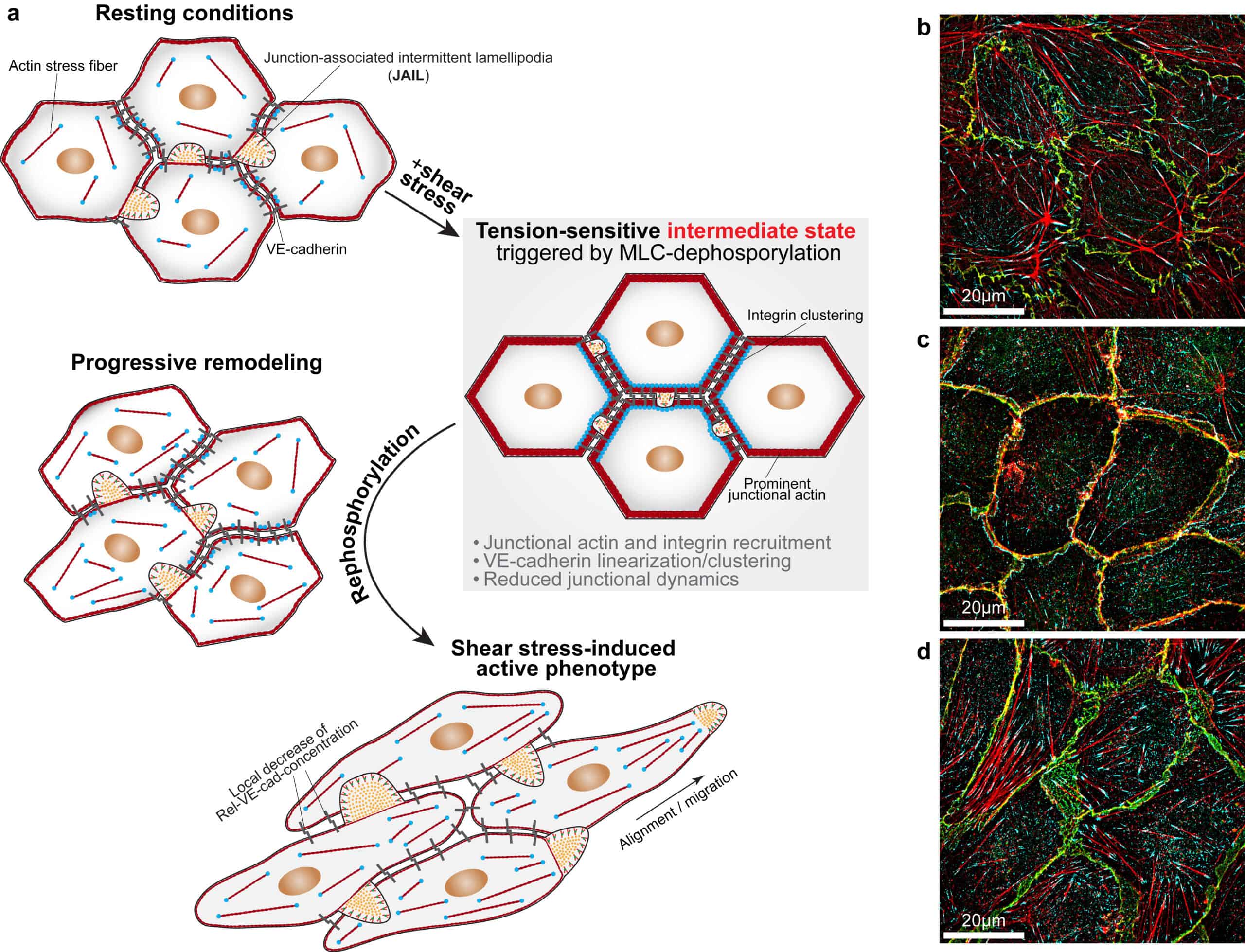
A tension-sensitive intermediate state mediates shear stress-induced endothelial remodeling and activation. (a) Scheme illustrating shear stress-induced endothelial remodeling from a resting to an active state: Resting endothelial cells (ECs) exhibited cell-cell junctions characterized by moderate junctional actin organization, high local VE-cadherin concentration and low junctional dynamics, as reflected by small junction-associated intermittent lamellipodia (JAIL). Upon 20 minutes of shear stress exposure, cells rapidly entered a transient, tension-sensitive intermediate state marked by myosin light chain (MLC) dephosphorylation, recruitment of prominent junctional actin and integrins, enhanced VE-cadherin clustering and linearization, increased barrier function and further suppression of JAIL formation. Consequently, cells adopted a homogeneous cobblestone-like morphology. Subsequent MLC-rephosphorylation initiated endothelial remodeling by dissolving junctional actin, promoting stress fiber reformation and VE-cadherin dilution, increasing JAIL-mediated junctional dynamics and weakening barrier function. These changes facilitated cell shape adaptation and migration, ultimately leading to the characteristic elongated phenotype aligned with the direction of flow. (b-d) Merged structured illumination microscopy images of ECs under (b) resting conditions, (c) during the tension-sensitive intermediate state and (d) after 4 hours of shear stress exposure. Cells were labelled for VE-cadherin (green), integrin b1 (cyan) and actin filaments (red). Panels (a-d) were assembled and modified from data published by Franz et al., 2026. Adapted under the terms of the Creative Commons Attribution 4.0 International License (CC BY 4.0).