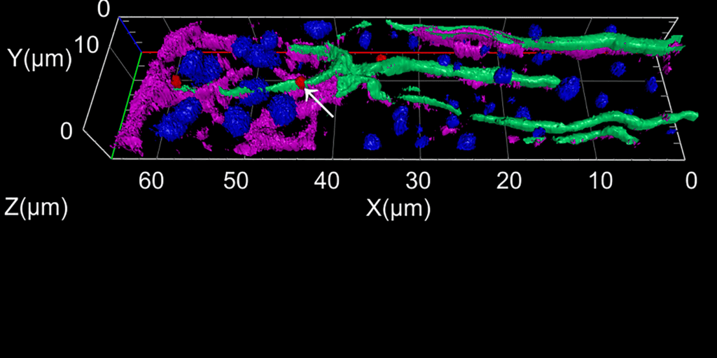
3D image of a GFAP (glial-fibrillary-acidic protein, green) -/GS (glutamine-synthetase, magneta)- positive, reactive Müller cell in the neurodegenerative retina. In situ hybridization for Tgfbr2 (transforming growth factor receptor 2, red) is shown. By combining in situ hybridization, immunofluorescence staining and 3D reconstruction, the research group of Prof. Barbara Braunger at the Institute of Neuroanatomy, University Hospital Hamburg-Eppendorf demonstrated for the first time that Tgfbr2 is expressed in reactive Müller cells (arrow) and in neurons upon retinal damage. Furthermore, GFAP reactivity in Müller cells indicates an ongoing neuroinflammatory process. Cell nuclei are stained DAPI (blue). ONL = outer nuclear layer, OPL = outer plexiform layer, INL = inner nuclear layer
Prof. Dr. med. Barbara Braunger, b.braunger@uke.de
Institute of Neuroanatomy, Universitätsklinikum Hamburg-Eppendorf
https://www.uke.de/kliniken-institute/institute/neuroanatomie/team/index.html